MAGNETIC RESONANCE IMAGING (MRI) at J|medical
There are multiple reasons that drive a doctor to request a magnetic resonance imaging scan for a patient; this is because it is an exam capable of providing very detailed images that can prove fundamental for the meticulous exploration of bones, muscles, organs, joints, and vessels. It is therefore able to offer a precise picture of the situation and the district the doctor has examined.
So what is magnetic resonance imaging? It is a non-invasive medical imaging technique that is not harmful to the patient, safe, and painless. It provides three-dimensional images (very detailed and sharp), not only of soft tissues (such as muscles, ligaments, tendons) but also of hard tissues (bones, cartilage) through the use of magnetic fields. It does not use ionizing radiation. The exam lasts an average of 20 minutes (variable based on the patient and the area to be examined).
What is it used for? It is a transversal exam across numerous fields of medicine, among which we recall traumatology, oncology, cardiology, orthopedics, gastroenterology.
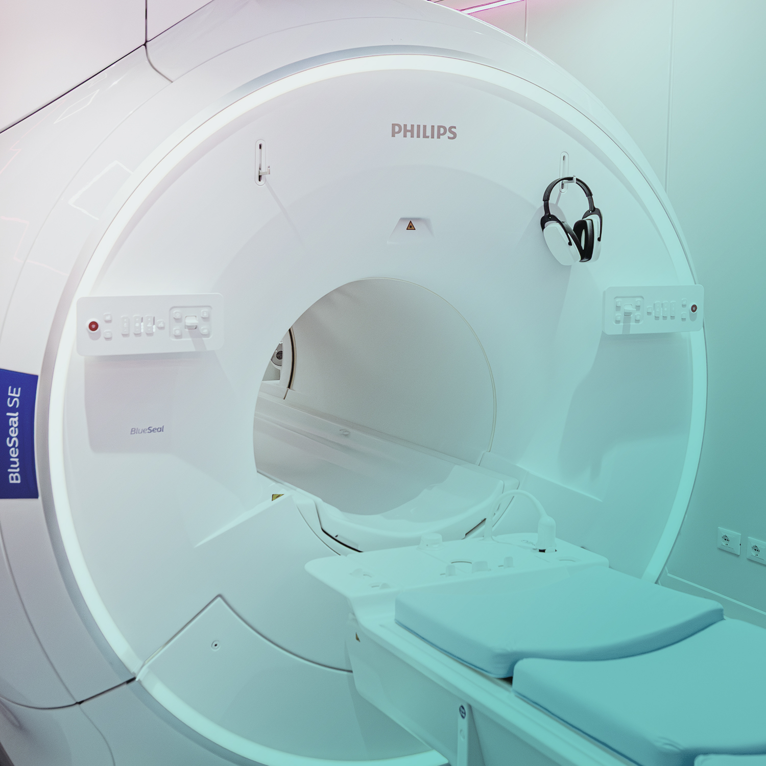
Is it possible to perform this exam at J|medical? Absolutely yes. At our center, it is possible to perform magnetic resonance imaging with a latest-generation device (equipped with a wide gantry, the space inside which the patient is positioned), which reduces the sensation of oppression. It is capable of producing high-quality images.
Numerous professional athletes and various sports clubs of excellence turn to J|medical for the execution of these diagnostic exams.
SERVICES AT J|medical:
Articular and Musculo-tendinous MRI. These are exams that can possibly also be performed with endo-articular contrast medium (Arthro-MRI). They are essential for diagnosing and evaluating a wide range of pathological conditions, such as musculo-tendinous lesions following sports trauma; inflammations; joint degenerations. Articular and musculo-tendinous MRIs guide treatment planning, helping doctors make important decisions on the therapy to adopt.
Shoulder Arthro-MRI represents the reference exam regarding the study of lesions affecting the rotator cuff and ligaments and fibro-cartilaginous structures in the shoulder joint, allowing the evaluation of damage caused by dislocations or other traumas.
Neurological MRI. These are exams that allow for obtaining very detailed images of the nervous system, with particular reference to the brain, the spinal cord, and nerve structures. They allow for a precise evaluation of pathological conditions, such as post-stroke brain damage, staging of lesions in multiple sclerosis, and identification of brain tumors, even small ones.
Internal MRI. Includes exams such as multiparametric prostate MRI and breast MRI. They often represent second-level exams capable of clarifying even very important diagnostic doubts. There are also particular investigation techniques with contrast media dedicated to the study of various organs, such as hepatospecific contrast for the liver and gadolinium, which allows for highlighting tissue alterations in the initial phase.
Recently at J|medical, the magnetic resonance has been updated through the innovative implementation of software that uses AI (artificial intelligence) for image processing. This translates into better image quality and a significant reduction in the execution time of the investigation.